



Lymphoma
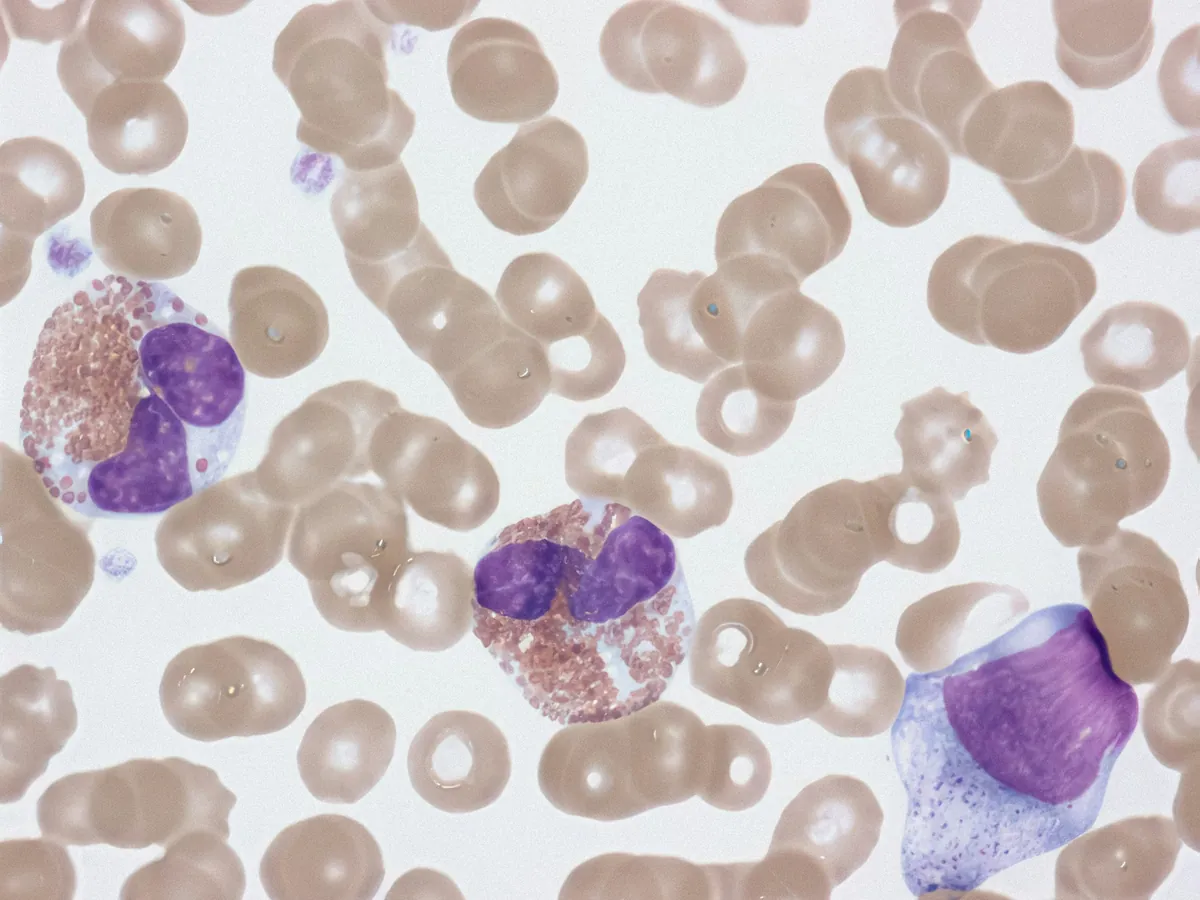




To diagnose. The only test that confirms lymphoma. A core or excisional biopsy is needed; a fine-needle sample is usually too small to name the subtype.
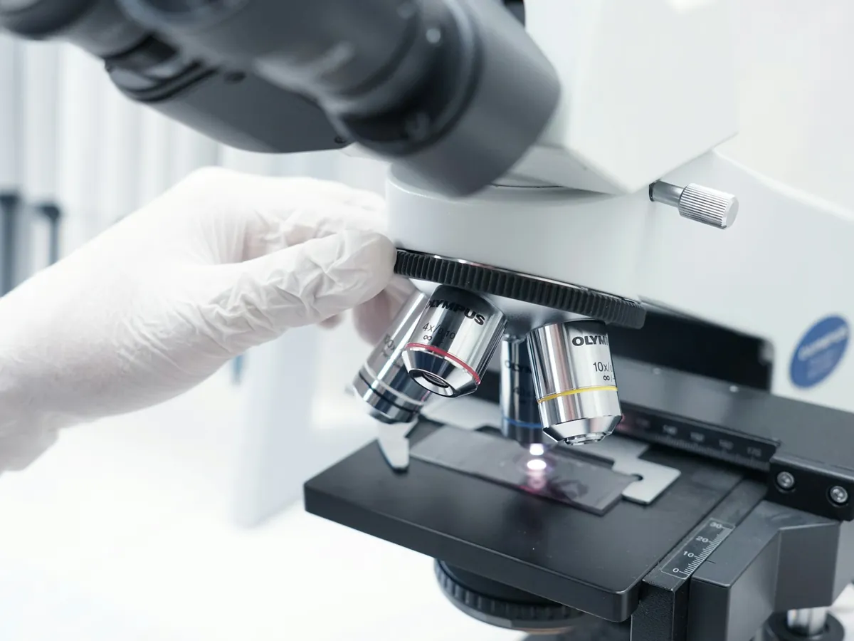
On the biopsy. Stains for proteins such as CD20 and CD30 that identify the subtype and the targets drugs can attack.
On the biopsy, blood or marrow. Reads surface markers on thousands of cells to show a single abnormal clone.
Aggressive B-cell lymphoma. Finds rearrangements of MYC, BCL2 and BCL6, which can change the treatment chosen.
Selected subtypes. Mutations that refine the diagnosis or point to a targeted drug.
At diagnosis and before every cycle. Shows whether the marrow is involved and whether counts are safe for the next dose.
At diagnosis and during treatment. Kidney and liver function, which decide drug doses.
At diagnosis. Rises with tumor burden, and is part of the IPI risk score.
Before the first cycle. Flags the risk of tumor lysis syndrome when treatment starts.
Some subtypes. A risk marker in follicular lymphoma and CLL/SLL.
Hodgkin lymphoma. Part of the risk assessment for early-stage Hodgkin lymphoma.
Before rituximab or similar drugs. These drugs can reactivate hidden hepatitis B. A positive result usually means preventive antiviral medicine.
Before treatment. Linked to some lymphomas; treating the virus can matter for the lymphoma too.
Before treatment. Changes the treatment plan and the infections to guard against.
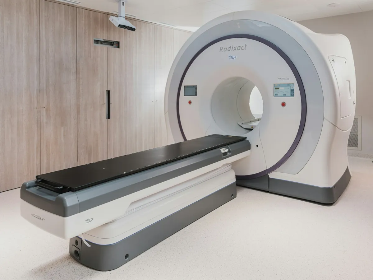
Staging, and after treatment. Shows where the lymphoma is and, afterwards, whether any remains active (the Deauville score).
When PET-CT does not settle it. Checks the marrow. Often skipped in Hodgkin lymphoma and DLBCL when PET-CT answers the question.
Before anthracyclines. Measures heart function before drugs such as doxorubicin, which can weaken it.
Before bleomycin. Bleomycin can scar the lungs; a baseline makes any change visible.
Before treatment. Chemotherapy and radiotherapy can harm a pregnancy. Fertility preservation is discussed at the same time.
When the risk to the brain and spine is high. Checks spinal fluid in the subtypes and stages that can spread there.
CLL/SLL, and after rituximab. Low antibody levels raise infection risk and can be replaced.
At diagnosis. Deficiency is common and easy to correct. Low levels are linked with worse outcomes in studies, though cause and effect are unproven.
Fevers, night sweats, weight and swollen nodes, dated, for the hematologist.
Every test in the workup, ticked off as it is done, with the result beside it.
Your stage, your risk score and the factors behind them, in your own words.
The questions worth asking before treatment starts, with space for the answers.
Nausea, fevers, fatigue and mouth sores, dated, so the team can adjust the next cycle.
Your treatment summary, follow-up schedule and late-effect checks, in one place.
The others

How tests are chosen, and how to use them · 2 guides · 76 min
Several causes, told apart by testing · 4 guides · 152 min
An immune disease that shows on the skin · 4 guides · 158 min
Oil, blocked pores, bacteria and hormones · 4 guides · 146 min
Pigment lost to an immune attack · 4 guides · 142 min




